You are here: Nature Science Photography – Image creation, Depth and Size – Visual image creation
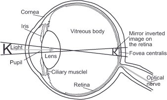
The encyclopedia defines „seeing“ as „the reception of light stimuli by the eyes and the perception of the information content of these optical stimuli in the brain.“ In humans and vertebrates, light rays are refracted by the cornea, lens and vitreous body, resulting in an inverted actual image on the retina. The optic nerve transmits the relevant action potentials from the retina to the cerebral cortex, where they are processed.
Living beings‘ physical reactions to light are approximately 1.5 billion years old. Its early version most likely helped creatures convert physical activity from night to day, and light-sensitive cells on the skin that perform this purpose may still be examined in primitive single-celled animals today. In a second phase, the photoreceptors were placed in small pits to protect them from stray light and increase the perception of moving shadows and potential danger. To shield these early eye pits from external things, translucent membranes gradually formed over them, becoming thicker in the center and laying the groundwork for the creation of a type of lens. The first of these lenses may have just functioned to magnify light, and it took several million years for them to produce truly useful images. It wasn’t until some 800 million years ago that eyes evolved, allowing living beings with various receptors to see both during the day and night. The eyes are essential to our vision today because they help the brain capture visual information. While the eyes resemble a camera in certain ways, they do more than just send a highly focused image to the brain; they also handle the first stage of the complex processing of acquired data.
The human eye, as we know it now, is a roughly spherical object around 2.5 cm in diameter. The dense tissue of the sclera protects it from the outside, allowing light to enter only through the small transparent area of the cornea. The gelatinous mass of the so-called vitreous humor occupies the majority of the eye’s interior, keeping the entire structure in shape and protecting the sensitive portions. The conjunctiva covers the cornea, which is the eye’s outermost functional unit. It refracts the incident light most strongly and, when combined with the lens, produces a sharp image. The next station inward is a tiny hollow filled with aqueous humor, which houses the iris. It is made up of thin connective tissue that contains the pigmented cells that give the eyes their various colors. However, this is just a means to an end because, aside from the pupil (also known as the pupil hole or iris diaphragm) in the middle, the iris must be completely lightproof. The retina, located in the back of the eye and responsible for reproducing the image seen, adjusts slowly to changes in luminance, so the iris serves as a protective diaphragm that closes quickly. It controls the pupil size between 2 and 8 millimeters, allowing it to reduce or increase the amount of incident light by two logarithmic units. Only after the iris makes an immediate adjustment do the sensory cells in the retina grow used to the new luminance. In addition to regulating light, the iris diaphragm is analogous to the camera aperture in that its constriction improves the depth of field during near vision.

The eyes are more than optical instruments. They perform the first stage of neurological processing of visual signals.
The trick of an eye mirror is required to look through the pupil into the eye, because the person observing’s head constantly creates a shadow. Only while photographing with light can we frequently get an unintended glance inside the eye. If the flash is too close to the lens’s axis and the pupil is wide open due to poor ambient lighting, the retina, which is well supplied with blood, appears as a red reflection in the image. Flash units can solve this by either constricting the pupil with a succession of pre-flashes that reflect little light back, or by unleashing the flash, offset from the exposure axis.
The lens is located immediately behind the iris. It is responsible for the eye’s adaptation to various object distances. For this reason, the ciliary muscle on the right and left sides of the eye contracts or relaxes, and this movement is transmitted to the lens via the zonula fibers, which changes curvature. If the object to be focused on is more than 6 meters away, the light rays fall nearly parallel to the retina, resulting in a sharp image. When the object gets closer, the picture plane changes behind the retina, and the rays no longer appear in parallel. To allow for near vision, the muscle contracts and, surprising enough, relaxes the zonula fibers, causing the lens to curve more sharply. The increased curvature also causes the light to be refracted more forcefully, and the image plane shifts forward sufficiently to allow the now sharp image to fall back onto the retina. This type of adjustment, known as accommodation, prevents muscle vibrations from being transmitted to the ocular system. The lens, like an onion, is constructed of layers. It grows throughout time as new cells adhere to its outer surface. Unfortunately, this development process has the unintended effect of eventually cutting off the flow of nutrients to the older cells on the inside, causing them to lose their suppleness. With age, the lens can no longer accommodate the optical system’s adaptation to varied distances, and glasses or contact lenses must compensate for this loss.
The interaction of the cornea, iris, pupil, and lens produces a sharp, tiny, and upside-down image of our surroundings on the inside of the eye and the retina that lines it, similar to a camera obscura. For a long time, it was thought that the brain understood the image put onto the retina as a whole using a type of „inner eye.“ However, contemporary research has demonstrated that visual perception is far more sophisticated.
Next The retina
Main Image creation, Depth and Size
If you found this post useful and want to support the continuation of my writing without intrusive advertising, please consider supporting. Your assistance goes towards helping make the content on this website even better. If you’d like to make a one-time ‘tip’ and buy me a coffee, I have a Ko-Fi page. Your support means a lot. Thank you!


 Since I started my first website in the year 2000, I’ve written and published ten books in the German language about photographing the amazing natural wonders of the American West, the details of our visual perception and its photography-related counterparts, and tried to shed some light on the immaterial concepts of quantum and chaos. Now all this material becomes freely accessible on this dedicated English website. I hope many of you find answers and inspiration there. My books are on