You are here: Nature Science Photography – Image creation, Depth and Size – Visual image creation
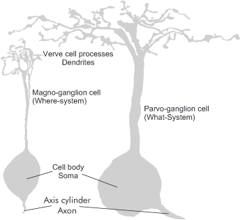
Even before the action potentials of the neurons triggered by the light stimulus leave the retina, an important division of information takes place. A little further above, it was already mentioned that the retina has two main genera of ganglion cells, the small magno ganglion cells and the large parvo ganglion cells. Both types are distributed throughout the retina and receive their input through the branches at the top, the dendrites. The more pronounced the dendrites are, the more photoreceptors they come into contact with. The number of these contacts is called the receptive field of the cell. Regardless of the location in the retina, the large magno ganglia always have larger receptive fields than the small parvo cells. The ganglion cells send their signals to the brain via the nerve output on their underside. The optic nerve, which leaves the eye at the so-called blind spot of the retina, sums up all of these fibers.

The distinction between the magno- and parvo ganglion cells is of such great importance because they establish two different perceptual channels, one of which is color-blind (i.e., uses only brightness values) and the other is color-sensitive: the where-system and the what-system. Both channels extend from the retina’s last layer to the higher brain areas. However, the separation of information is no longer quite strict, because as processing becomes more specialized, it becomes apparent that different visual attributes are processed in combination (Gegenfurtner, Kiper & Fenstemaker 1996). For instance, studies have demonstrated that we can perceive shape and motion in objects solely based on color (i.e., those with equivalent or isoluminant brightness values) (Gegenfurtner & Hawken 1996).
From the study of monkeys whose magno- or parvocellular layers in the corpus geniculatum laterale (see next section) were temporarily switched off experimentally, we know that both systems process the information provided by their progenitor cells according to different aspects. Animals in which the magno layers were disrupted showed marked limitations in motion vision, while those with inhibition of the parvo layers showed deficits in color and depth perception (Schiller, Logothetis & Charles 1990). These findings are supported by experiments with people who suffered a spatially confined stroke. If they suffered lesions in the branch of the where-system, they exhibited various apraxias, disturbances in the visual information processing that underlie the control of motor functions. Damage in the branch of the what-system resulted in agnosia (disturbance of object recognition), prosopagnosia (disturbance in the ability to recognize faces) or central achromatopsia (loss of color perception). These studies also demonstrated that the what-system further subdivides into a shape system, which employs both brightness and color to delineate object outlines, and a low-resolution color system, responsible for determining surface color. The following list provides a detailed summary of the properties of the two main channels.
Where-system

Color: is colorblind

Contrast: has high contrast sensitivity

Speed: works at high speed, but tires quickly. It therefore only performs a superficial analysis of the scene

Resolution: is low because the ganglion cells are each connected to all three photoreceptors present
What-system

Color: processes color information

Contrast: requires a larger difference threshold between light and dark

Speed: runs at a slower speed and is therefore more enduring. This is because it is used to develop a scene in detail
resolution: is higher by a factor of two to three because the ganglion cells are connected to only one or two photoreceptors. However, the what channel itself is further subdivided into a shape system, which uses brightness and color information to recognize the shapes of objects, and a low-resolution color system, which describes the surface colors.
Naturally, the question arises as to why the visual system has partitioned and parallelized perception in this manner, and why the properties of the two channels must differ so significantly. A look at evolutionary history provides the answers. The where-system is ancient and found in all mammalian species. It is sufficient for them to orient themselves spatially in their environment, to distinguish objects and, most importantly, to detect movement because what is moving is either food or a predator, i.e., important. To fulfill these requirements, it is unnecessary to perceive colors or to recognize objects very precisely. This only became important with the rise of higher mammals, including primates. Instead of developing a completely new visual system for them, evolution retained the existing one and simply added a second layer on top, which provided the now-needed abilities. While not perfect, this method is the easiest and most resource-efficient. And according to the last premise, evolution always acts.
We can further develop the resource conservation argument to justify separate information processing. Processing data that describes the same thing simultaneously and at the same location is particularly effective and efficient. There is a natural separation between an object’s shape, color, and position in space or movement. Under this separation, the brain does not need to connect potentially distant areas, which would be a waste of scarce resources, and can specialize each individual area in the necessary way.
Thus, the main task of the neural network in the retina is to channel the output signals of the photoreceptors according to certain features. Color, shape, direction of motion, and speed are the main keywords here. We extract data from an image of a red car driving down a busy street based on these features: Speed and direction of the drive and all further movements, the shapes and lines of the different objects transport the where-system, the distinguishable wavelengths of the incident light, from which the color impression becomes, flow in the what-system. – We know from the computer that such abstract, vector-oriented data, respectively data shrunk to their characteristics, require less storage space and processing capacity than the total number of all points that make up an image. Additionally, the brain can only manage the accumulation of large amounts of information in a sufficient amount of time by separating and processing the visually perceived data in parallel. Surprisingly, we also find the same approach in the field of high-definition digital television (HDTV) and computer graphics. There, information about the shape and color of an object is handled separately from that about its position and direction of motion.
Next Fourth step: Forwarding and filtering
Main Image creation, Depth and Size
Previous Second step – Beginning of information processing
If you found this post useful and want to support the continuation of my writing without intrusive advertising, please consider supporting. Your assistance goes towards helping make the content on this website even better. If you’d like to make a one-time ‘tip’ and buy me a coffee, I have a Ko-Fi page. Your support means a lot. Thank you!


 Since I started my first website in the year 2000, I’ve written and published ten books in the German language about photographing the amazing natural wonders of the American West, the details of our visual perception and its photography-related counterparts, and tried to shed some light on the immaterial concepts of quantum and chaos. Now all this material becomes freely accessible on this dedicated English website. I hope many of you find answers and inspiration there. My books are on