You are here: Nature Science Photography – Image creation, Depth and Size – Visual image creation
Light transports visual information, and the optics of the eye produce a two-dimensional representation of the surroundings and objects on the retina. Particular sensors known as photoreceptors read the enclosed energy potential. At the current level of evolution, each of our retinas contains roughly 120 million highly specialized sensory cells that convert light into electrical signals and notify the visual system about the strength and chromaticity of the incident spectrum. We discriminate between around 110 million rod cells, named for their distinctive forms, and approximately 6 million cone cells.
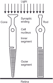
The basic structure of both receptor types consists of three parts: the outer segment, the inner segment, and the synaptic body. To receive as little reflected light as possible, they position themselves „upside down“ on the retina. The outer section consists of approximately 1,000 stacked membrane discs, each containing the photochemically active pigment. The fundamental key to vision is a combination of the big protein opsin and the little light-sensitive molecule retinal, a vitamin A derivative. Because they absorb light, they have a distinct color: a somewhat dark, opaque purple known as visual purple. When exposed to light, the pigment bleaches and becomes opaque white, rendering it worthless for the visual process. The inner section is responsible for replacing it. It regenerates wasted molecules, incorporates them into new membrane discs, and transports them to the outer segment, where they gradually reach the tip. In addition, the inner section houses the cell nucleus and the mitochondria (the cell’s „power plants“), which sustain energy metabolism through protein synthesis. Finally, through the synaptic body, the receptor connects to the downstream retinal cells.

Figure 3: Cross section of the two human photoreceptor types
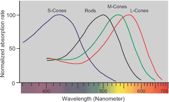
All rod cells contain the photochemically active pigment rhodopsin, which makes them sensitive to wavelengths ranging from 440 to 620 nanometers (green-yellow). Each cone cell contains one of five distinct iodopsin pigments, which cover the spectral range from 400 nm (blue) to 700 nm (red), with a sensitivity maximum at 580 nm (yellow). The opsin’s genetic code is responsible for limiting the wavelength range. According to this assignment, they are also known as S cones (short wave, blue), M cones (medium wave, yellow), and L cones (long-wave, red).

Let’s take a closer look at pigment bleaching, because it’s vital to the overall visual process. In darkness, the electrical potential difference between the inside and exterior of the cell is -30 millivolts due to a continual input of sodium ions. During this stage, the synapse permanently discharges messenger chemicals, thereby inhibiting the retinal cells from further processing them. When exposed to light, the photochemically active pigment breaks down into its parts, which are the protein opsin and the dye retinal. The opsin, which is now free, changes how permeable the cell membrane is through a chain of enzymes. The cell experiences hyperpolarization when the conduction channels close, preventing the afterflow of sodium ions, thereby providing potential equalization and lowering the membrane potential to its resting value of -70 millivolts. Since the receptor no longer emits messenger chemicals and no longer inhibits the retina’s downstream cells, they continue to deliver an excitation signal, resulting in the perception of brightness and color.
This is how vision works: light modifies the photopigments, which causes an electrochemical reaction that influences the activity of the synaptic connection and sends an impulse to the nervous system.
Why are we so sensitive to the limited spectrum between 400 and 700 nanometers? – Well, the wavelength range below 380 nm (ultraviolet) is so intense that it would swiftly damage the photopigments in our eyes and, after a little longer amount of time, turn the eye lens yellow. Some bird and insect species have developed UV sensitivity, but they die before the light can cause any substantial damage. Larger mammals, like humans, have longer life spans and must adapt their visual systems to these harmful effects. On the other side of the spectrum, wavelengths greater than 780 nm (infrared) are mostly thermal radiation, providing little information on the nature of things. A face appears as a hot iron skeleton on infrared film, so this long-wavelength radiation provides little information about the world under daylight. So our vision ignores the extremes of the spectrum and instead focuses on the middle region, which interacts most powerfully and differentially with matter and informs us the most about the world.
Next Second step – Beginning of information processing
Main Image creation, Depth and Size
Previous The retina
If you found this post useful and want to support the continuation of my writing without intrusive advertising, please consider supporting. Your assistance goes towards helping make the content on this website even better. If you’d like to make a one-time ‘tip’ and buy me a coffee, I have a Ko-Fi page. Your support means a lot. Thank you!


 Since I started my first website in the year 2000, I’ve written and published ten books in the German language about photographing the amazing natural wonders of the American West, the details of our visual perception and its photography-related counterparts, and tried to shed some light on the immaterial concepts of quantum and chaos. Now all this material becomes freely accessible on this dedicated English website. I hope many of you find answers and inspiration there. My books are on