

You are here: Nature Science Photography – Contrast – Contrast perception – The dynamic range of the visual system
However, our eyes contain two types of photoreceptors, and one of their fascinating characteristics is their varying sensitivities. By analogy with photography, the retina has two types of film: a sensitive SW film (the rods) and a less sensitive color film (the cones). The different sensitivities of both are due to the pigment inherent in each. Rhodopsin decays at low light levels, whereas iodopsin needs more energy to do so. For this reason, rod cells are active primarily at low illumination levels. For instance, they function best in the evening and at night. The cone cells, on the other hand, work almost exclusively during the day and enable us to see in bright light. The rod and cone apparatus adapt to changes in brightness, known as light adaptation and dark adaptation, respectively. Regeneration issues and related chemical processes cause both adaptations. Within adaptation, we can distinguish three main states:
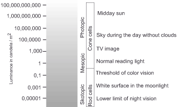
Scotopic vision(night vision), for which the rod receptors are responsible at luminances between 3*10-6 cd/m² and 3*10-2 cd/m² (=0.000003 to 0.03 cd/m²) (4 log10 units).
Mesopic vision(twilight vision) marking the transition between the two main adaptation stages at luminances between 3*10-2 cd/m² and 3*100 cd/m² to 3*101 cd/m² (=0.03 and 3 cd/m² to 30 cd/m², respectively), in which both cones and rods are active (1 log10 unit each for rods and cones).
Photopic vision(day vision), which the cone receptors perform at luminances between 3*100/3*101 and 3*106 cd/m² (= 3/30 cd/m2 to 3 000 000 cd/m²) (6 log10 units). The specified limits are fluid and individually different.
So, when we go from photopic rod vision to scotopic cone vision, we see a big change in adaptation. We also see a lot of smaller changes because the receptors become more sensitive during these two stages.
We can imagine the first case approximately as follows: Let’s say you go from the brightness of a summer day to your dark basement. Perhaps your goal is to grab your bike and head to the outdoor swimming pool. At first it is too dark for you to see anything, and you probably bump into a piece of furniture standing around. However, gradually, your sensitivity adapts to the altered general brightness, allowing you to see details at first. However, as time passes, you begin to notice more and more colorless details in the otherwise dark environment. During this adaptation phase, a light stimulus, such as the red glowing emergency stop switch next to the boiler room door, will gradually appear brighter and brighter to you.
At the level of the receptors, the following happens: In the very first moment of darkness, you see nothing at all, because the previously acting exposure has greatly reduced the sensitivity of rods and cones. After this moment of shock, both receptor types use the opportunity to regenerate their pigment. This happens fastest in the cones, and their slightly increased sensitivity brings out the initially hazy outlines. After five to ten minutes, when the cones cannot increase their sensitivity any further, the rods have regenerated enough to begin contributing their part. As they adapt, their characteristics determine our perception, enabling us to perceive more details over time, despite their near-colorlessness.
If you go outside again after some time, the great brightness will initially blind you for a moment. During this brief period, the rods bleach most of their rhodopsin supply, which can only partially regenerate as long as the photopic conditions remain unchanged. They are virtually saturated, and without pigment to decay, the receptors naturally emit no signal. However, the return of sufficient light triggers the cones, supplying us with the necessary information for color perception.
The second case, adaptation within one of the two main states, occurs when the ambient brightness does not change to the complete opposite but still changes noticeably. This occurs, for instance, when we transition from bright daylight into the shadowed area of a large tree. Now, we can regenerate pigment proportionate to the brightness change, thereby increasing the light sensitivity of the receptors.
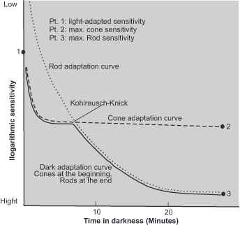
To indicate the sensitivity of the receptors within the adaptation states, we can measure the brightness of a currently perceivable light stimulus and plot it as a curve across the time axis. The resulting graph is called an adaptation curve (figure 13). Its vertical axis indicates the brightness of the light stimulus. Since the range of our sensitivity is very large, we use a logarithmic scale here. Values at the upper end represent large stimulus brightness and mean that our sensitivity is low. Those at the lower end represent low brightness, but conversely, high sensitivity. The horizontal axis indicates time in darkness in minutes. Read from left to right, the figure tells us that when we have just moved from light to dark, a strong brightness stimulus is needed to be perceived. During the following minutes, the receptors increase their sensitivity, and the brightness threshold for perception decreases first very quickly and then more slowly. In this phase, during the first five to ten minutes, we can definitely still indicate the color of the perceived light stimulus, and this is a sure sign that our cone cells are still active. Then the curve changes direction a little, and the sensitivity again increases abruptly. After reaching this prominent point, also known as the Kohlrausch-Knick, the visual system responds to weaker light stimuli, but we are no longer able to distinguish their color because the system has now shifted to the rods. The curve continues to decrease until it reaches the bottom after approximately 30 minutes, at which point it becomes straight. After this extended period of darkness, we are able to detect a weak light stimulus that corresponds to a single candle from a distance of 16 km!
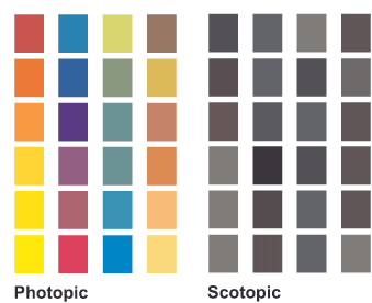
The flexible system of adaptation ensures that our eyes always work with the right sensitivity, just as in photography we adjust film speed to the ambient brightness. Similarly, we trade resolution and sharpness for sensitivity in our visual perception. Because, as the previous section has demonstrated, the fovea centralis, the site of sharpest vision, lacks rod receptors, and their density in the remaining part of the retina decreases towards the edges of the retina. Moreover, in scotopic vision, the visual system allows color to „fall ‚down the back.'“ – Recording technology no longer requires this sacrifice, as it did when the most sensitive films were always black and white.
But the change from scotopic to photopic vision (from rods to cones) has another consequence than the mere lowering or raising of the general sensitivity. The section on the perception of brightness and color addressed the spectral sensitivity of the receptors.
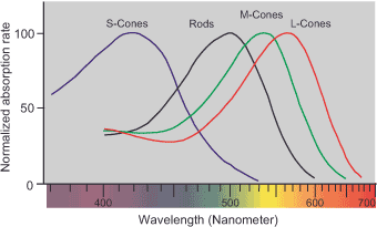
Figure 14: Normalized absorption spectra of rod and cone cells. According to data from Bowmaker, J.K., Dartnall, H.J.A.: Visual pigments of rods and cones in a human retina.
Journal of Physiology Nr. 298: S. 501-511 (1980)
Both rods and cones respond differently to different wavelength regions of the spectrum. The rods respond best to the short-wavelength blue region, while the cones respond best to the more long-wavelength red region. Therefore, under photopic conditions, if we view an object through the cones, it will appear brighter to us than an objectively equally bright blue object. Under scotopic conditions, the situation reverses. The Purkinje phenomenon, named after its discoverer, refers to this change in the perceived brightness of different colors.
Next Lateral inhibition
Main Contrast
Previous The response range of the photoreceptors
If you found this post useful and want to support the continuation of my writing without intrusive advertising, please consider supporting. Your assistance goes towards helping make the content on this website even better. If you’d like to make a one-time ‘tip’ and buy me a coffee, I have a Ko-Fi page. Your support means a lot. Thank you!
 Since I started my first website in the year 2000, I’ve written and published ten books in the German language about photographing the amazing natural wonders of the American West, the details of our visual perception and its photography-related counterparts, and tried to shed some light on the immaterial concepts of quantum and chaos. Now all this material becomes freely accessible on this dedicated English website. I hope many of you find answers and inspiration there. My books are on