You are here: Nature Science Photography – Image creation, Depth and Size – Visual image creation
So we now understand how light becomes nerve impulses. Something with which the neurological system can function. However, this is just the start of the problems, as these impulses are not merely transmitted and then observed. Instead, the visual system processes the input from the first stage. An example demonstrates exactly what it accomplishes. Consider figure 5 (Mach’s stripes). It depicts a series of areas with varying gray colors that lack color gradation. Nevertheless, you will notice that the individual stripes appear as gradients from light to dark, with the brightness difference increasing towards the boundaries. This phenomenon is known as Mach’s stripes after its discoverer, the physicist and philosopher Ernst Mach (1838-1916), and it has long been unknown how it occurs.

Figure 5: Mach’s stripes
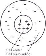
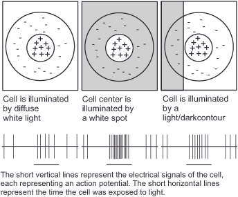
We have Stephen Kuffler (1913-1980) to thank for the realization that vision is more than simply transporting the retinal image to a location in the brain where it is viewed. His research demonstrated that vision is a process of information processing, as he found the first and most essential phase of this cascade in the 1950s. He observed the activity of retinal ganglion cells and discovered that he could cause them to „fire“ with little bits of light. Of course, it was long known that the eye responds to light, but Kuffler took a methodical approach and discovered that the smaller the irritating point of light, the better the cells responded. Based on the fact that large dots were less effective than small ones, he concluded that ganglion cells were not only excited by light falling on the centers of their receptive fields (the retinal area they covered), but also inhibited when light fell on the immediate vicinity of the centers.

Figure 6: A retinal ganglion cell in center/surround organization. The plus and minus signs indicate which areas of its receptive field react to light and how.
This cell organization, known as center/surround, is critical for stimulus processing in the nervous system because it makes the cells sensitive to interruptions in the light patterns in the retinal image (the edges and border areas of the objects) while being insensitive to changes in the absolute amount of light or its gradual change, both of which are less important. Many visual sensations, including brightness, color, motion, and spatial depth, are dependent on center/surround organization.
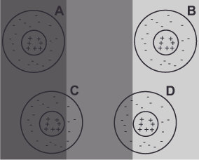
With the center/surround organization, Mach’s stripes can be explained on the basis of figure 7 (Explanation Mach’s stripes) as follows: Cell A is least excited by the darkest stripe in comparison. The receptive field of cell B, on the other hand, falls on the brightest stripe, which excites it the most. The exciting receptive center of cell C falls entirely within the darkest first stripe, whereas its inhibiting receptive environment lies partially within the slightly brighter second stripe. For this reason, the environment generates an inhibitory response that causes the cell to „see“ a darker stripe as a result than those cells whose receptive fields lie entirely within the same stripe (for example, cell A). We see the opposite phenomenon in cell D. Its exciting light-responsive center lies entirely in the third brightest stripe, while its inhibiting environment lies partly in the darker middle stripe. Here, too, the environment generates an inhibitory response, which this time makes the cell „see“ a brighter stripe than cell B.


The contrast enhancement (that the inner edges appear darker and the outer edges lighter) at the borders between the individual stripes in figure 5 (Mach’s stripes) is thus due to the competition between cells whose receptive fields lie entirely within one stripe and those whose receptive fields lie to some extent in the respective other stripe. The perceived brightness gradients within the stripes stem from the fact that as the distance to the edge increases, the cells are inhibited less and less, and eventually not at all, by their surroundings, resulting in a fine staircase formation.
Still missing is the rationale for the emergence of the center/surround organization. It makes a lot of sense, as it is economical for the visual system to process objects based on light pattern interruptions. This allows it to encode only the parts of the image that undergo changes, rather than the entire image. Edges and border areas are the only important information that the apparatus in our heads needs to construct the shapes and forms of things in our environment. It is unnecessary to define brightness and color at every single point of, for example, a solid red object. Instead, it is quite sufficient to do this everywhere where something changes. And this is the case at an edge or boundary. This significantly reduces the amount of information required for transmission and processing.
We can see exactly how much by comparing two graphics in .tif or .jpeg format. Let’s assume that the image shows a red circle in the middle of a green area in 10×15 cm format. In .tif there are 1715 KB. In .jpeg only 13 KB – 132x less. The reduction is due to the fact that .jpeg, just like the visual system, only defines those pixels where something changes. The file only contains the border position and color on the inside or outside. The image processing program automatically fills in the pixels in between.
This reduction in the amount of information is eminently important for the nervous system in general, because for a nerve cell to fire, energy is needed and the body must use this raw material as sparingly as possible. – Keep in mind that the brain has a particularly high demand for oxygen and energy. It makes up only about 2% of the body’s mass but consumes about 20% of the oxygen and more than 25% of the glucose. So the fewer nerve cells that are active, the better it is for the organism.
Next Third step – Categorization of information
Main Image creation, Depth and Size
Previous The photoreceptors in general
If you found this post useful and want to support the continuation of my writing without intrusive advertising, please consider supporting. Your assistance goes towards helping make the content on this website even better. If you’d like to make a one-time ‘tip’ and buy me a coffee, I have a Ko-Fi page. Your support means a lot. Thank you!


 Since I started my first website in the year 2000, I’ve written and published ten books in the German language about photographing the amazing natural wonders of the American West, the details of our visual perception and its photography-related counterparts, and tried to shed some light on the immaterial concepts of quantum and chaos. Now all this material becomes freely accessible on this dedicated English website. I hope many of you find answers and inspiration there. My books are on