You are here: Nature Science Photography – Image creation, Depth and Size – Visual image creation
On their way to the higher processing centers of the brain, the data divided into the where and the what channels now pass the junction of the visual pathway (optic chiasm) a few centimeters behind the eyes. Each retina is divided vertically into a left and a right half (quasi inside and outside). At the junction, the optic nerve fibers of these two halves switch sides to travel to their corresponding cerebral hemisphere. Thus, the right halves of each retina are represented in the right cerebral hemisphere, and the left halves of each retina are represented in the left cerebral hemisphere. This type of data division allows the visual apparatus to compare the images of both eyes and this lays the foundation for the perception of spatial depth.
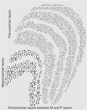
The corpus geniculatum laterale (CGL), also known as the knee bump, is the first higher processing station in the diencephalon thalamus. This is where about 90% of the optic nerve fibers terminate. The remaining 10% run past the CGL or through it without switching and terminate in the hypothalamus, area praetectalis and superior colliculi. Their information is not for vision but for reflex head and eye movements, pupillary reflex, day-night rhythm, etc. While the retina provides some input, the cerebral cortex, thalamus, and other CGL neurons provide much more. The primary visual cortex receives this combined information as visual radiation. As a result, the CGL functions as a powerful switching station for the visual system, acting as a gatekeeper by evaluating and eliminating various stimuli. It manifests the division of information between the two ganglion classes in two anatomically clearly distinguishable areas.
At the bottom are the two magnocellular layers, to which the signals of the large magno-ganglion cells run. They have conspicuously large cells and are developmentally the oldest, which is why we share them with all other mammalian species. In them, the branch of the where pathway continues, which is why they are responsible for the perception of movement, spatial depth and three-dimensionality, position, figure-ground separation, and general organization of a visual scene. Although they are colorblind, they are highly sensitive to contrast and work very fast.
The four parvocellular layers, which receive their input from the smaller parvo-ganglionic cells, are located at the top. Their cells are more finely structured and they are found only in developmentally relatively young primate species, which include us humans. With them, the branch of the what pathway continues, and so they are responsible for our detailed object perception, including faces. They respond highly selectively to color information, are less sensitive to contrast, and operate more slowly than the where cells.
In the approximately 1.5 million cells of the CGL, an initial coarse and still unconscious perception manifests itself in the form of lines, shapes and colors, which are initially sorted according to their importance. The CGL is thus a filter that saves the brain from having to process the whole variety of visual impressions. The visual data found to be important enough are allowed to pass through the gatekeeper CGL and reach the primary visual cortex

Figure 9: Section through the corpus geniculatum laterale (CGL), the lateral geniculate body in the thalamus
Next Excursus – Brain and nerve cells
Main Image creation, Depth and Size
Previous Third step – Categorization of information
If you found this post useful and want to support the continuation of my writing without intrusive advertising, please consider supporting. Your assistance goes towards helping make the content on this website even better. If you’d like to make a one-time ‘tip’ and buy me a coffee, I have a Ko-Fi page. Your support means a lot. Thank you!


 Since I started my first website in the year 2000, I’ve written and published ten books in the German language about photographing the amazing natural wonders of the American West, the details of our visual perception and its photography-related counterparts, and tried to shed some light on the immaterial concepts of quantum and chaos. Now all this material becomes freely accessible on this dedicated English website. I hope many of you find answers and inspiration there. My books are on