You are here: Nature Science Photography – Image creation, Depth and Size – Visual image creation
After this brief digression, we return to our pursuit of data processing in the visual system. The corpus geniculatum laterale sends its neurological excitation patterns to the
primary visual cortex, which is made up of about 200 million cells and is about 3 mm thick and the size of a credit card. It is located at the back of the two cerebral hemispheres. It is developmentally younger than the CGL, and it seems only logical that its sophisticated data analysis represents an evolution of the CGL’s primitive but still useful unconscious vision.

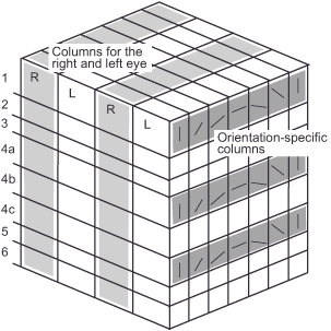
Figure 13: Schematic structure of the visual cortex. Layers 1-3 forward data to brain areas in the “where pathway” or the “what pathway”. Layers 4a-4c receive data from the CGL Layers 5-6 forward data back to the CGL/thalamus
Neurobiological research on the cat’s visual cortex reveals the division of the visual cortex into parallel layers, with blocks running perpendicularly through them, one for the left eye and one for the right eye alternately. This structure, akin to a crossword puzzle, represents a kind of map where each box processes signals from a single distinct area of the retina. However, the representation of all areas is not equal, as the zone of sharpest vision, known as the fovea centralis, occupies only 0.01% of the retinal surface but accounts for 8% of the neurons in the primary visual cortex. Therefore, the high concentration of rod and cone cells and their connections in the retina, along with the disproportionately large area of the visual cortex for further processing, contribute to this area’s high visual acuity.
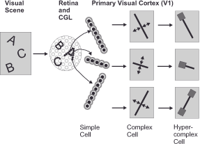
The real peculiarity of many cells in the complicated structure of the primary visual cortex was recognized by David Hubel and Thorsten Wiesel, both students of the pioneer Stephen Kuffler, six years after his groundbreaking discovery of center/surround organization, in 1958. After spending hours trying to excite a single cell in a cat’s visual cortex with circular patterns of various sizes and recording extraordinarily sporadic responses, they discovered, more or less by accident, that these responses were not due to the small round stimuli but occurred whenever they moved the test glass into the projection apparatus or pulled it out. Hubel writes, „Except for a headstand, we tried just about everything to stimulate the cells to fire“ and from this one can see that both must have been already a little frustrated (Hubel 1995). When the glass shadow fell exactly horizontally on the retina, the cell reacted very precisely. All other orientations elicited only silence. This was a dam break, and Hubel and Wiesel tested numerous other cells with line patterns oriented in all conceivable directions. The finding: Within the vertical blocks of area V1 of the primary visual cortex, we find so-called simple cells (oriented cells) in each layer, which only react to a very specific spatial orientation (horizontal, vertical, diagonal and the gradations in between) or direction of movement (from left to right, from top to bottom, etc.) of the occurring visual stimuli with an electrical impulse. Assuming normal development, according to the research results, all possible orientations and directions of movement are equally present in the number of neurons. However, they respond best to stimuli that come in the form of a narrow stripe (Hubel & Wiesel 1959). Hubel and Wiesel’s discovery suggests that the next step in visual information processing after center/surround organization, which serves to detect all the basic transitions and edges of a visual scene, is that of sorting these edges and boundaries by direction. To achieve this, the cells of area V1 of the visual cortex integrate the data of individual CGL ganglion cell groups. The direction to which an oriented cell is sensitive depends on the orientation of the receptive fields of the center/surround cells that feed it.
Further research on functionally higher layers of the visual cortex revealed cells that respond to extended, occluded or interrupted contours, corners or curvatures (Hubel & Wiesel 1962, 1965). The so-called complex cells, also known as extended contour cells or hypercomplex cells, process the pre-digested data from simple cells grouped together. From processing stage to processing stage, cells become more specialized and respond to larger visual fields. Thus, the visual system is able to construct the complex objects that make up our environment from the random individual edges at the level of the center/surround cells. Quite as if we were making a picture by simply connecting individual points.
David Hubel and Thorsten Wiesel had recognized that the ganglion cells in the retina, CGL (center/surround), and their relatives in the visual cortex (simple cells and complex cells) form a hierarchy of information processing, and that vision is just that: an active process of information processing and not merely passive image perception!

However, the work of these two scientists does not end there. In the 1970s, they researched the development or non-development of the cells and structures of the visual system. They found out that in kittens, for example, the darkening of an eye for two or three months after the first days of life leads to permanent blindness due to an irreversible underdevelopment of nerve cells in the visual apparatus. Adult animals, on the other hand, quickly regain normal vision after the same procedure. Further animal experiments, where young animals grew up under strict control of their visual environment (e.g., only vertical black and white stripes or vertical contours on one eye and horizontal ones on the other), revealed the loss of neurons in the primary visual cortex sensitive to other orientations. Consequently, the mere failure to perceive visual patterns and movements during a certain period of development, when the brain exhibits a special plasticity, leads to permanent maldevelopment (Hubel 1978).
The brain must first train all its functions, which essentially means that the neuronal circuits must adapt to the stimuli presented. The brain changes constantly and as long as the organism lives, however, the first phase of life is particularly important.
And even in the adult brain, learning processes and external influences can alter the visual system’s basic structure. We stimulated the nerve cells of the visual cortex of experimental animals with weak electrical currents to provoke these effects. The energy flux activated many cells in an area measuring only about 1/10 mm, initially changing the transmission of stimuli between them for a short time, but then permanently by repeated activation over a few hours. The change that could be tracked because it could be seen from the outside by measuring blood flow caused orientation-sensitive cells in the visual cortex to move in an area a few millimeters across. Real visual stimuli also trigger increased electrical activity in the brain and are likely to cause similar changes, although not as rapidly as the actually meaningless artificial stimuli. David Hubel and Thorsten Wiesel received the Nobel Prize in Physiology/Medicine in 1981 for their pioneering discoveries.
The shapes and patterns we see influence the shape of the primary visual cortex and thus directly our constructed image of the world – so perceiving means shaping!
Comparing the visual apparatus of humans to that of experimental animals establishes that our environment directly influences the neuronal level of our perception, thereby affecting it at its core: Frequently observed visual patterns reflect in a disproportionately large number of specially sensitized neurons and, most likely, also manifest in fixed synaptic circuitry, leading us to subconsciously favor these patterns in our selection. The repeated viewing of certain images or patterns, therefore, directly influences our style. This is exactly why we may ask ourselves with Galen Rowell, “ … how much of the visual ability of our greatest photographers already existed as a fixed neuronal circuitry in their heads before they picked up a camera for the first time“ (Rowell 1993).
Next Sixth step – Generation of impressions
Main Image creation, Depth and Size
Previous Excursus – Brain and nerve cells
If you found this post useful and want to support the continuation of my writing without intrusive advertising, please consider supporting. Your assistance goes towards helping make the content on this website even better. If you’d like to make a one-time ‘tip’ and buy me a coffee, I have a Ko-Fi page. Your support means a lot. Thank you!


 Since I started my first website in the year 2000, I’ve written and published ten books in the German language about photographing the amazing natural wonders of the American West, the details of our visual perception and its photography-related counterparts, and tried to shed some light on the immaterial concepts of quantum and chaos. Now all this material becomes freely accessible on this dedicated English website. I hope many of you find answers and inspiration there. My books are on