You are here: Nature Science Photography – Image creation, Depth and Size – Visual image creation
After this stay at the lowest level of the visual system, it is time to see where the nerve impulses emitted by the photoreceptors travel to and what happens to them there. Of course they travel to the brain; where else! But what is the brain actually? Does the brain simply reflect our environment through a vast cluster of cells, or does it function as a structured organ that organizes our perception and much more in specific subareas? Prior to discussing details, we must first understand the big picture.
The brain, in its essence, consists of an unimaginably large number of about 200 billion nerve cells, or neurons. They produce the brain’s input and output signals, those weak electrical impulses that make our perception and thinking possible in the first place.

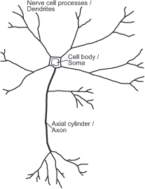
Figure 10: Schematic representation of a nerve cell
According to their function, neuronal processes, cell bodies and axial cylinders can be roughly divided into input, processing and output. The dendrites, for example, sum the output signals of the surrounding neurons caused by light stimulation of a retinal sensory cell in the form of an electrical potential. If a certain threshold is exceeded in the soma, a short electrical impulse is generated, which is transmitted to the neighboring cells via the axon; we say that the cell „fires“. Contact points known as synapses transmit this impulse. Synapses sit on the ramifications of dendrites. Depending on the type and state of the synapse, an incoming impulse causes a potential increase or decrease of varying strength in the target neuron, as well as a feedback process that further increases or decreases the activity of all neurons involved.
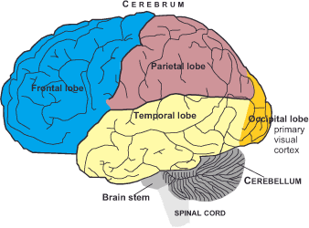
In the course of evolution, the brain has developed from a center controlling only the life support systems to the consciousness-giving control center of our body. Outwardly distinguishable are the cerebrum, which is particularly conspicuous with about 80 % of the brain mass, the cerebellum, which occupies the lower area of the rear part, and the brain stem, which lies in front of it and merges with the spinal cord.

The cerebrum is the most highly developed area. Functional centers such as movement, sensory perception, hearing, sight, smell, language and memory are assigned to it. Anatomically, it divides into the left and right cerebral hemispheres. There are several parts to the cerebrum, which primarily coordinates muscle movements. The brainstem controls the basic functions of blood circulation, heartbeat and lung activity, as well as reflexes such as yawning, coughing, sneezing and vomiting. Not visible from the outside, two other parts lie inside the brain: the diencephalon, consisting of the thalamus (sensory and motor functions), hypothalamus (hormonal system) and epithalamus (biological clock), and the midbrain, which controls eye movements, among other things.
The brain – a big pile of complexly interconnected neurons that create our environment as an intelligible reality.
For the process of visual perception, the cerebrum is most important. The two halves of the cerebrum, from front to back, distinguish the following sections, which correspond to the right and left cerebral hemispheres: The frontal lobe, occupying the broad frontal area, which is responsible for attention and the onset of muscular activity. The temporal lobe, which is located to the side and is responsible for processing language and conceptual thinking. The parietal lobe, which is responsible for sensory perception, spatial-visual processes and body orientation, and the occipital lobe, which is located at the back of the brain and is the home of the primary visual cortex.

Next Fifth step – The visual cortex and beyond
Main Image creation, Depth and Size
Previous Fourth step: Forwarding and filtering
If you found this post useful and want to support the continuation of my writing without intrusive advertising, please consider supporting. Your assistance goes towards helping make the content on this website even better. If you’d like to make a one-time ‘tip’ and buy me a coffee, I have a Ko-Fi page. Your support means a lot. Thank you!


 Since I started my first website in the year 2000, I’ve written and published ten books in the German language about photographing the amazing natural wonders of the American West, the details of our visual perception and its photography-related counterparts, and tried to shed some light on the immaterial concepts of quantum and chaos. Now all this material becomes freely accessible on this dedicated English website. I hope many of you find answers and inspiration there. My books are on