You are here: Nature Science Photography – Lightness and color – The perception of lightness and color
In evolutionary terms, the retina is an externally displaced portion of the brain. It is only 1/10 millimeter thick and has around 200 million closely packed, highly specialized nerve cells. The upside-down image of our environment falls on it. The retina is a curved plane that corresponds to the curvature of the eyeball, giving it the advantage of being the same distance from the lens at all points and providing a sharp image everywhere. In addition, the curvature provides for the same proportion of the image scale regardless of the angle of incidence of the light.

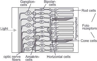
The structure of the retina is notable for stacking its functional layers so that light only reaches photosensitive cone and rod cells after passing through the neuronal cells above them. This approach involves inserting a film with the photographically active side facing outward, which decreases contrast-reducing stray light. It is achievable without hazard because the neural plexus on top does not move, and our perception filters out such silent impulses from our conscious view.
From back to front, the photoreceptors are followed by horizontal cells, then bipolar cells and amacrine cells, and finally ganglion cells. Each of these neuron types occurs in different varieties and fulfills other tasks in addition to the following basic functions. For example, there are over a dozen different types of amacrine cells, as well as two major genera of ganglion cells: small magno cells and giant parvo cells. Bipolar cells receive input signals directly from photoreceptors and many of them have connections to ganglion cells. Horizontal cells transport data between individual receptors, while amacrine cells do the same for individual bipolar cells. This type of linkage allows for feedback (lateral inhibition) as well as the grouping of specific receptors or bipolar cells
Next Determining the physiological input stage – The photoreceptors in general
Main Lightness and Color
Previous Determining the physiological input stage – The eye
If you found this post useful and want to support the continuation of my writing without intrusive advertising, please consider supporting. Your assistance goes towards helping make the content on this website even better. If you’d like to make a one-time ‘tip’ and buy me a coffee, I have a Ko-Fi page. Your support means a lot. Thank you!


 Since I started my first website in the year 2000, I’ve written and published ten books in the German language about photographing the amazing natural wonders of the American West, the details of our visual perception and its photography-related counterparts, and tried to shed some light on the immaterial concepts of quantum and chaos. Now all this material becomes freely accessible on this dedicated English website. I hope many of you find answers and inspiration there. My books are on