

You are here: Nature Science Photography – Lightness and color – The perception of lightness and color
The British physicist Thomas Young was the first to predict, as early as 1801, in his three-color theory of vision, that three different types of receptors are responsible for our color vision, providing three different types of information. Hermann von Helmholtz, who further advanced Young’s research, assumed that these receptors are not equally sensitive over the entire range in which they respond, but are more sensitive in one and less sensitive in another. He developed three hypothetical absorption curves for the photoreceptors, based on the assumption that they differed in terms of spectrum and stimulus response quality at that time. Figure 8 (Helmholtz resonance curves) shows the curves assumed by Helmholtz. Each of these curves spans a broad range and exhibits optimal response within a specific wavelength range. The curve on the far left describes a receptor that responds best in the short-wavelength region; the one in the middle responds to the mid-wavelength region of the spectrum; and the one on the right shows the assumed behavior of a receptor with the best response in the long-wavelength region. Following this gradation, we call these cone receptors S receptor (for short wavelength), M receptor (for medium wavelength), and L receptor (for long wavelength).
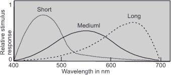
A color stimulus excites either one, two, or all three types of receptors and is converted into a specific signal pattern. A stimulus such as the remission curve of the green object in figure 2 (remission curve) would excite the M and L receptors most strongly and the S receptors only a little. This excitation pattern is the basis for the perception of green that emerges after further processing steps. Since the excitation patterns depend on the exact shape of the absorption curves, it is critical that we determine them as precisely as possible.
To do this, we make use of modern microspectrophotometry, which allows us to determine wavelength by wavelength the amount of light that each receptor absorbs. A weak light pulse is sent to a precisely defined spot on the retina and precise measurement technology is used to determine how much of it is reflected. The result of this analysis is that only three different absorption spectra could be determined, and Thomas Young received a late but nevertheless undoubted confirmation: Our retina does indeed have three different cone receptor species that are responsible for color perception based on the spectral sensitivity of their photochemically active pigments. The relationship between their sensitivity and the wavelength of light is expressed in an absorption curve unique to each receptor type, shown in
figure 9 (Absorption spectra). The higher this curve rises, the more pigment bleaching occurs at the respective wavelength and the stronger the receptor’s output signal.
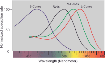
The S cones (for short wavelength) respond to the fairly narrow range of the spectrum between 400 nm and 520 nm (violet, blue and blue-green) and are most sensitive at a wavelength of about 435 nm (blue-violet). The M cones (for medium wavelength) respond in the wide range between 450 nm and 660 nm (blue, blue-green, green, and yellow) and have a sensitivity peak at 530 nm (green). The L cones (for long wavelength) cover an even slightly larger portion of the spectrum, as they are also sensitive to red-orange with a range of 460 nm to 700 nm. Their maximum sensitivity is around 565 nm in the green-yellow range. The fact that the individual cone types respond differently to the different wavelength ranges is due to the fact that they are filled with iodopsin pigments, which are genetically different from each other. In contrast, rod cells all contain the photochemically active pigment rhodopsin and are thus sensitive to the wavelength range between 440 nm and 620 nm (green-yellow).
From the absorption curves, we can understand what happens in the cone apparatus when we stimulate it with different spectra of light. This will explain why we perceive the two spectrally differently composed light stimuli of figure 3 (intensity distribution curves C and D) as identical color impressions.
In the case of the monochromatic yellow with a wavelength of 570 nm (figure 3D), we get a strong signal in the long-wavelength channel and a moderate one in the mid-wavelength. In the case of the polychromatic yellow mixed from green (530 nm) and red (650 nm) (figure 3 C), the green excites the M cones strongly and the L cones less strongly, whereas the red excites only the L cones. In sum, this raises the response of the L cones above that of the M cones. Thus, the combined response is identical to the one we found when stimulated with monochromatic yellow light at 570 nm, which is why we perceive yellow in both cases. In this manner, we can explore all other potential combinations and observe that they consistently result in the same outcome: the same stimulus response for each of the three cone types, leading to an identical perception of color. So the receptors do not care how they are stimulated. We will perceive the same color impression if only their output quantities are the same, which we can guarantee with only three basic colors.
The cone cells in the retina sort the spectrum roughly according to the intensities of the different wavelengths and pass it on as encoded stimulus patterns.
Next First processing stage – Categorization of information
Main Lightness and Color
Previous Determining the physiological input stage – The photoreceptors in general
If you found this post useful and want to support the continuation of my writing without intrusive advertising, please consider supporting. Your assistance goes towards helping make the content on this website even better. If you’d like to make a one-time ‘tip’ and buy me a coffee, I have a Ko-Fi page. Your support means a lot. Thank you!
 Since I started my first website in the year 2000, I’ve written and published ten books in the German language about photographing the amazing natural wonders of the American West, the details of our visual perception and its photography-related counterparts, and tried to shed some light on the immaterial concepts of quantum and chaos. Now all this material becomes freely accessible on this dedicated English website. I hope many of you find answers and inspiration there. My books are on